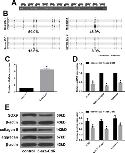
Figure 6. MiR-494 expression is regulated by promoter methylation status.

(A) Representative results of MSP analysis of miR-494 in human NP tissue from mild and severe IDD groups. M, methylated primers; U, unmethylated primers. (B) Representative results of BSP of miR-494 methylation in human NP tissue from mild and severe IDD groups. Black and white circles represent methylated and unmethylated CpG sites, respectively. (C–E) Mild IDD NP cells were treated with 5 μM 5-aza-CdR for 3 days, and untreatedcells were used as a control. (C) MiR-494 expression in mild IDD NP cells, as determined by qRT-PCR. U6 was used as an internal control. (D, E) Transcript (D) and protein (E) levels of SOX9, type II collagen, and aggrecan in mild IDD NP cells, as determined by qRT-PCR and western blotting, respectively. β-actin was used as a control. Data represent mean ± SD. *P < 0.05 vs. control group.
