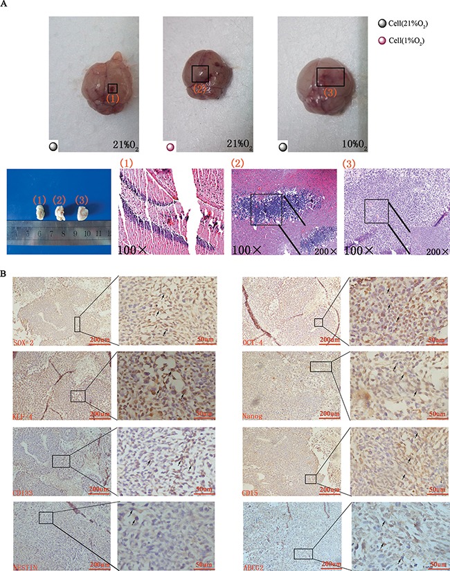
Figure 6. Tumor formation and immunohistochemical staining in vivo.

(A) Gross anatomy indicated no tumor formed when normoxia-cultured CD133−CD15−NESTIN− cells were injected into the mice raised under normoxia (1 represented group 2 and 3), but tumor formed when hypoxia-cultured CD133−CD15−NESTIN− cells were injected (2 represented group 1) or normoxia-cultured CD133−CD15−NESTIN− cells were injected into the mice but raised under hypoxia (3 represented group 4). Similar results were demonstrated via HE staining. (B) Immunohistochemical staining demonstrated the increased expression of stem cell markers in tumor samples.
