
Figure 1. Representative analytical proteome map of rectal cancer (RC).
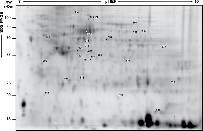
Proteins were resolved by isoelectrofocusing over the pI 3-10, followed by 8–16% gradient second dimension. Numbered spots indicate the differentially expressed spots in RC biopsies of ‘TRG 1-2′ versus either ‘TRG 3′ or ‘TRG 4′. Identified proteins are listed in Table 2.