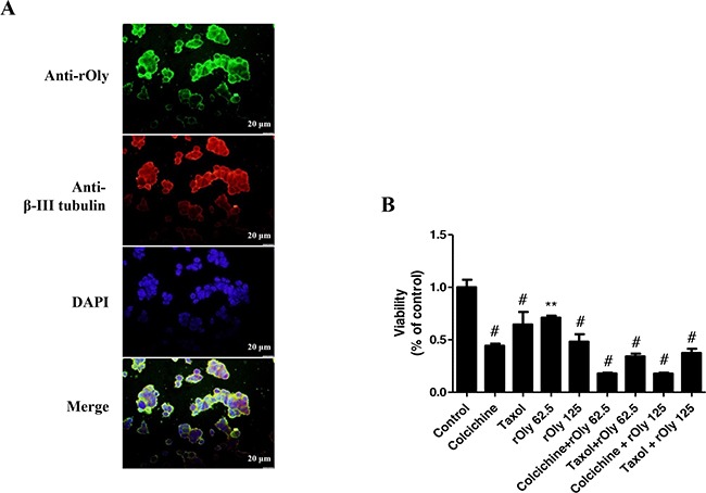
Figure 4. Recombinant ostreolysin (rOly) and tubulin-inhibiting agents decrease HCT116 cell viability.

A. Immunofluorescence of β-III tubulin and rOly in HCT116 cells. Cells were treated for 90 min with 125 μg ml−1 rOly and labelled with specific anti-rOly (green) and anti-β-III tubulin (red) antibodies. Nuclei were counterstained with DAPI. Scale bars = 20 μm. This is one representative section out of three independent experiments. B. HCT116 cells were grown overnight in DMEM and then subjected to different treatments as described in Materials and Methods. Cell viability was estimated by MTT assay. #P < 0.001, **P < 0.01 as compared to control, n = 6 (one-way ANOVA–Dunnett's test). Data are presented as means ± SEM of one independent experiment out of three.
