
Figure 3. Apoptosis of K562 cells induced by Stel B.
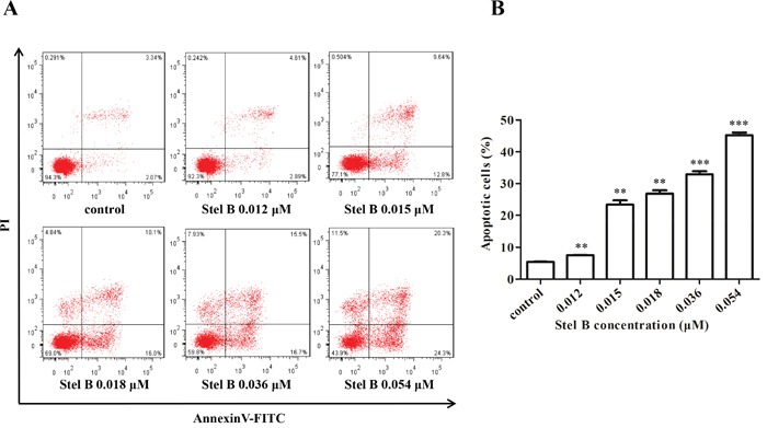
(A) Flow cytometric analysis of cell apoptosis with Annexin V-FITC/PI double staining. K562 cells were harvested 48 h after treatment by indicated concentrations of Stel B, stained with Annexin V-FITC/PI and subjected to flow cytometry analysis. (B) Quantification of the apoptotic cells in both early and late stage. Data represent mean± SD of three independent experiments. *: p < 0.01, ***: p < 0.001, compared with control.