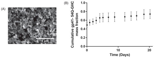
Figure 1.
gp41-54Q-GHC antigen release kinetics from polyanhydride nanoparticles and structural characterization from scanning electron photomicrographs. Panel A shows a scanning electron photomicrograph of gp41-54Q-GHC-loaded 20:80 CPTEG:CPH nanoparticles. Scale bar: 1 μm. Panel B shows the cumulative fraction of gp41-54Q-GHC released (•) from the 20:80 CPTEG:CPH nanoparticles. Error bars represent standard error of the mean; results are representative of three independent experiments.

