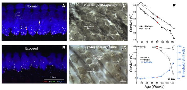
Fig. 1. Noise-induced and age-related loss of synapses and SGNs.
Evaluating synaptopathy by triple-staining cochlear whole mounts for a pre-synaptic marker (CtBP2-red), a postsynaptic marker (GluA2-green) and a hair cell marker (Myosin VIIa-blue). Confocal z-stacks in the IHC area from a control (A) and a noise-exposed mouse (B), 2 wks post exposure. Light micrographs of osmium-stained plastic sections from noise-exposed ears, 2 wks (C) or 2 yrs (D) post exposure. Exposure in B and D was 8–16 kHz, 2 h, 100 dB SPL, delivered at 16 wk to CBA/CaJ mice. (E) In aging ears from the same inbred strain, synaptic counts at IHCs decrease steadily from 4 to 144 wks and parallel ganglion cell loss follows whereas, (F) threshold loss begins comparatively later and accelerates beyond 80 wks, mirrored by accelerating loss of OHCs. IHC loss is trivial at any age. Red symbols flag 80 wk data points for all measurements. After Kujawa and Liberman 2006, 2009; Sergeyenko et al. (2013).

