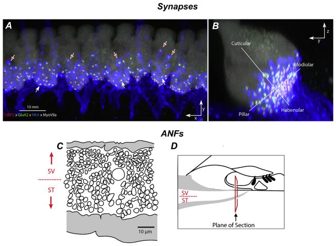
Fig. 5. Gradients in synaptic and afferent fiber morphology.
IHC synapses in confocal z-stacks, acquired in the x-y plane (A) and re-projected into the y-z (B) plane. A Pre- and post-synaptic elements in the IHC area are counted in cochlear whole mounts quadruple-immunostained for CtBP2 (red), GluA2 (green), NaK ATPase (blue), and myosin VIIa (white). B Size gradients in pre- and post-synaptic elements are quantified according to location along habenular-cuticular and modiolar-pillar axes (Liberman et al., 2015). (C) Tracing of peripheral axons from a cross section through the osseous spiral lamina (OSL; D) in a normal cat shows the SR-based gradient from thin (low-SR) to thick (high-SR) fibers (Kawase and Liberman, 1992).

