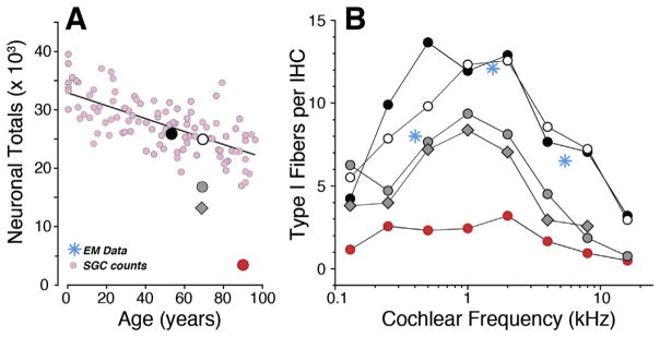
Fig. 6. Cochlear de-afferentation in human temporal bones.
Cochlear de-afferentation is seen in human temporal bones as a function of age (A) and cochlear location (B) in cases with no hair cell loss and no explicit otologic disease. The small pink symbols (A) are estimated total SGC counts from archival sections (Makary et al., 2011); the five large symbols (A) are the estimated total peripheral axon counts from the same five cases shown in B. Counts of cochlear nerve terminals per IHC (B) in 5 normal aging temporal bones (55–89 yrs) with no history of otologic disease (Viana et al., 2015). Blue symbols are counts of synapses per IHC from an electron microscopic study of a normal middle-aged human (Nadol, 1988).

