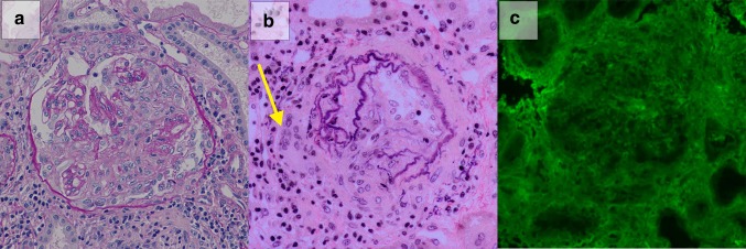
Fig. 2.
Renal biopsy specimen included 30 glomeruli, including 5 cellular crescents and 6 fibrocellular crescents. a Representative glomerulus containing cellular crescents with necrotizing lesion is shown (Periodic acid Schiff staining ×200). b Small artery exhibited granulomatous reaction with multinucleated giant cell formation (A arrow) and the destruction of elastic fibers (Elastica van Gieson staining ×200). c There was no staining of the IgG on immunofluorescence staining (×200)

