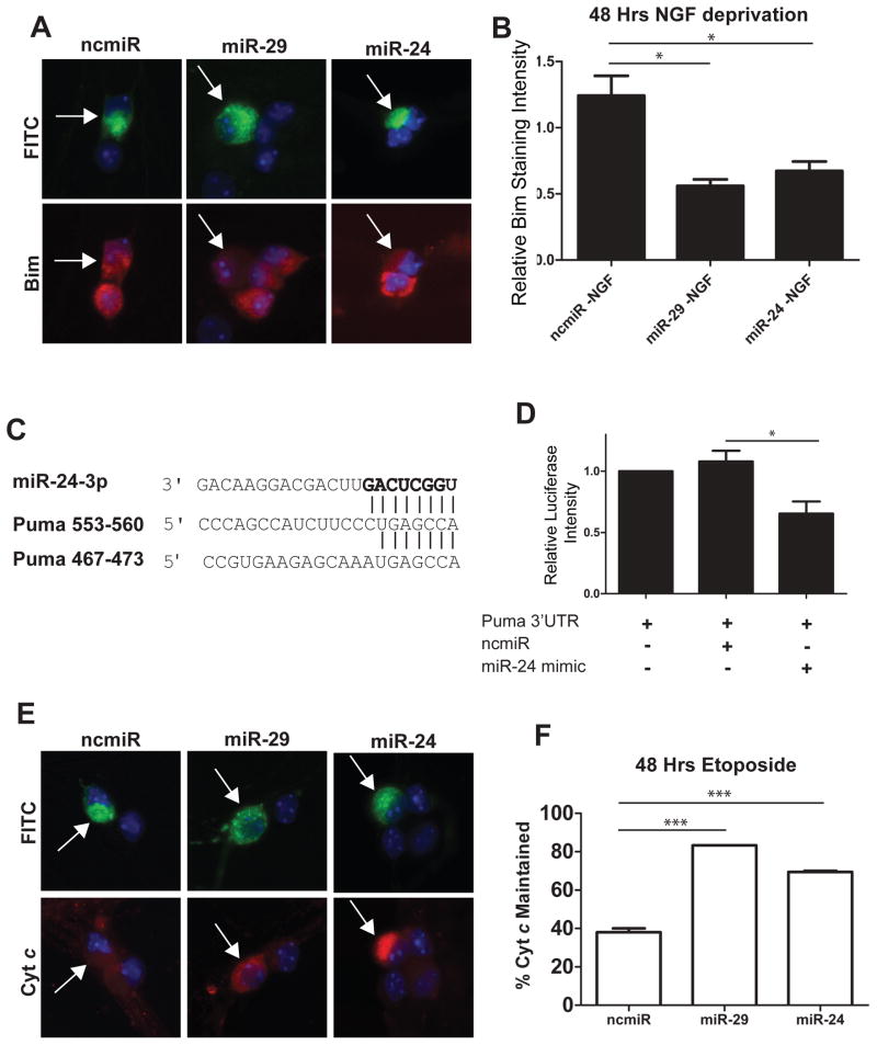
Figure 5.
Overexpression of miR-29 or miR-24 is sufficient to inhibit the induction of Bim and Puma in young sympathetic neurons. A) Representative images of Bim staining in neurons. Neurons were injected at 3 DIV with mimics to miR-29, miR-24, or a negative control, along with FITC-Dextran (Green) to mark injected cells. At 5 DIV neurons were deprived of NGF and after 48 hours of NGF deprivation, neurons were fixed and stained for Bim (red). B) Quantification of normalized Bim staining intensity. Bim staining intensity was measured, and values for injected cells were normalized to NGF-deprived, mock injected neurons. Data presented as mean intensity ± SEM of 3 independent experiments (*=P>0.05). C) Sequence and alignment of miR-24 seed sequence with two putative miR-24 target sites in Puma 3′UTR. D) Luciferase activity was measured 48 hours after transfection in HEK293T cells transfected with reporter plasmids containing the Puma 3′UTR fused to a firefly luciferase gene. Plasmids were transfected either alone or with 100 nM mimics of miR-24 or negative control. Expression was normalized by measuring the ratio of firefly to renilla luciferase. Values are plotted relative to vector alone and represent mean ± SEM of 3 independent experiments (*=P<0.05). E) Representative images of cyt c staining in neurons injected with mimics to miR-29, miR-24, or negative control miR (ncmiR) and treated with 20 μM etoposide. Green indicates injected cells (arrows). F) Quantification of cyt c release in young neurons injected with mimics to miR-29, miR-24, or negative control after 48 hours of etoposide treatment. Data are plotted as mean ± SEM of 3 independent experiments (***=P<0.001).

