Figure 1.

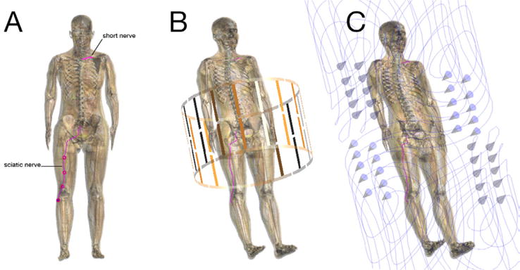
Simulation setup showing the anatomical model with highlighted nerve trajectories (A), inside the RF coil (B), and inside the gradient coil (the arrows indicate the orientation of the current flow in the coil) (C).

Simulation setup showing the anatomical model with highlighted nerve trajectories (A), inside the RF coil (B), and inside the gradient coil (the arrows indicate the orientation of the current flow in the coil) (C).