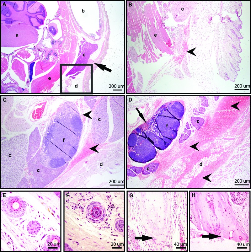
Figure 7.
Tissue sections stained with hematoxylin and eosin showed histologic changes associated with phlebotomy via (A–D ) the facial vein, (E and F) tail vein incision, or (G and H) tail tip amputation. (A) Transverse section of the head including the facial vein (arrow) and showing no significant histologic findings from a mouse with a single attempt at facial vein puncture. (B) Transverse section of the head, showing an enlarged version of the boxed region in A, from a mouse with a single attempt at facial vein puncture showing mild hemorrhage (arrowhead). (C) Transverse section of the head showing moderate hemorrhage in the subcutaneous tissue and surrounding the mandibular lymph node (arrowheads). (D) Transverse section of head showing extensive hemorrhage around the parotid gland, brown fat, and mandibular lymph node (arrow heads), with RBC in lymph node sinuses (arrow) . (E) Cross section of midtail with no significant findings in a TI mouse. (F) Cross section of mid-tail in a mouse from the TI group showing mild neutrophilic infiltrate in the deep dermis of cross section. (G) Longitudinal section of the tail tip showing an intact vertebra (arrow) with fibrin and mild neutrophilic infiltrate in a mouse from the TA group. (H) Longitudinal section of the tail tip showing the last caudal vertebra transected (arrow) with fibrin and moderate neutrophilic infiltrate in a mouse from the TA group. a, brainstem; b, external ear canal; c, salivary gland; d, adipose tissue; e, skeletal muscle; f, lymph node.

