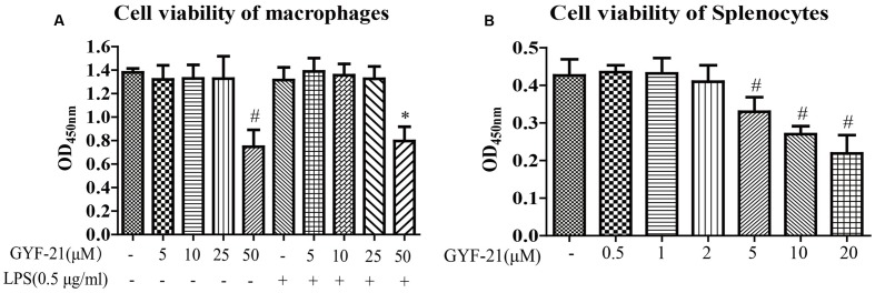
FIGURE 2.
Cytotoxicity of GYF-21 on microglia and splenocytes. BV-2 cells (A) and splenocytes (B) were treated with various concentrations of GYF-21 for 24 h and cell viability was determined with CCK-8. Data are representative of three independent experiments. #P < 0.05 vs. Vehicle; ∗P < 0.05 vs. LPS.

