
Figure 1.
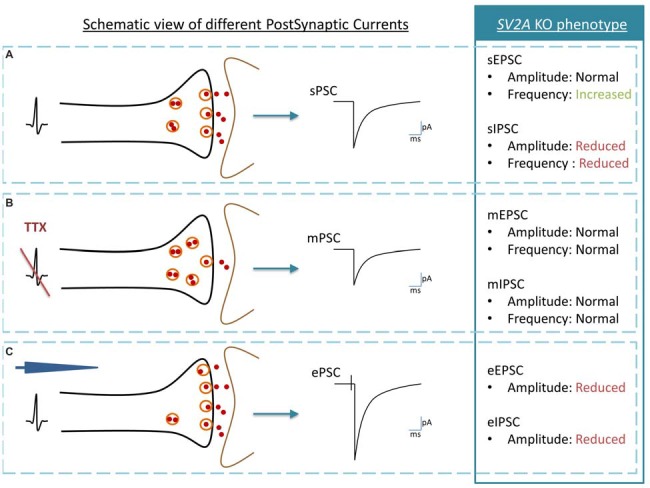
Schematic view of post synaptic currents (PSC) and summary of Synaptic vesicle proteins 2A (SV2A) KO phenotype. (A) Spontaneous PSC or sPSC: an action potential in the presynaptic neuron (black) induces a sPSC in the postsynaptic neuron (brown). (B) Miniature PSC or mPSC: the generation of action potentials is blocked by Tetrodotoxin (TTX). If SVs exocytosis is functional, a single vesicle is release by the presynaptic neuron (black) and induces a mPSC in the postsynaptic neuron (brown). The mPSC reveals thus the “molecular machinery” of SV exocytosis while sPSC also reveals the link between vesicle exocytosis and the pre-synaptic action potential. (C) Evoked PSC or evoked postsynaptic currents (ePSC): an action potential is induced with an electrode (blue) in the presynaptic neuron (black). The postsynaptic neuron (gray) responds by producing an ePSC. In comparison to the sPSC, the ePSC recruits the maximal possibilities of SVs exocytosis. The analysis of SV2A KO phenotype is telling us that: (1) in physiological conditions, inhibitory PSC are affected negatively by the SV2A absence, both in amplitude and in frequency although excitatory PSC are modified only through increased frequency; (2) the absence of SV2A does not interfere with the molecular mechanism of the SV exocytosis process; and (3) when the synapse is pushed at its maximum of activity, both excitatory and inhibitory PSC are decreased in amplitude.