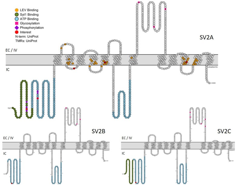
Figure 4.
Primary structures of SV2 proteins. In green: Synaptotagmin 1 binding domain. In blue: nucleotide binding domains. In orange: residues implicated in LEV binding. Purple lozenge: phosphorylated residues. Pink square: glycosylated residues. In red: residues of particular interest. EC, Extracellular; IV, Intravesicular; IC, Intracellular. (This figure was built using http://wlab.ethz.ch/protter/start/).

