Figure 3.
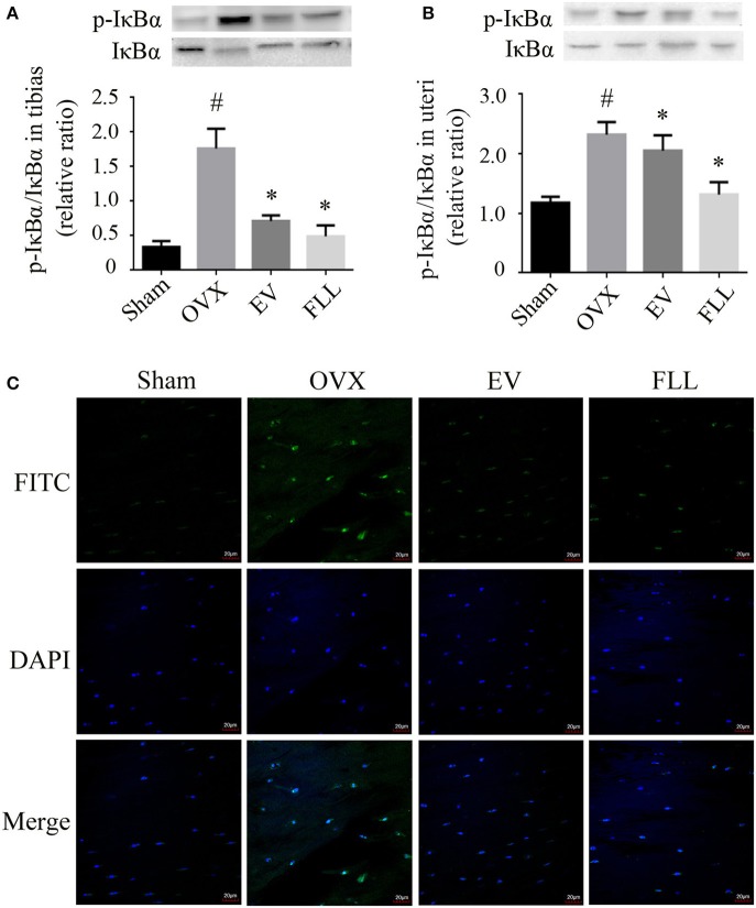
The representative western blot images and their analysis showed that FLL treatment decreased IκBα and p-IκBα expression in the tibias (A) and uteri (B) of OVX rats (n = 9). In addition, confocal microcopy of immunofluorescence staining (C; original magnification, × 60) showed that FLL blocked NF-κB-p65 nuclear translocation in the femurs of OVX rats. The green color represents NF-κB-p65 staining, the blue color represents nuclei staining, and the cyan (greenish-blue) color represents nuclear translocation. #p < 0.05 with Sham group, *p < 0.05 compared with OVX group.

