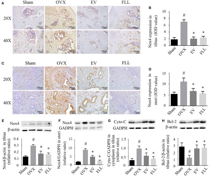
Figure 4.
The representative images of immunohistochemical staining (A–D; sections were counterstained with hematoxylin; original magnification, × 20), and western blot assays (E,F) showed that FLL treatment decreased Nox4 expression in tibias and uteri of OVX rats (n = 9). In addition, FLL treatment also decreased cytochrome C (Cyto-C; G) and increased Bcl-2 expression (H) in the tibias of OVX rats (n = 9). Data are presented as mean ± SD. IOD denotes integrated optical density of interested areas. #p < 0.05 with Sham group, *p < 0.05 compared with OVX group.

