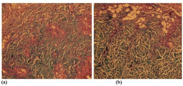
Fig. 1:
Microscopic view of open cutaneous wounds: (a) control group, collagen fibers are lesser than the experimental honey twice daily group in this photomicrograph. (b) Experimental honey twice daily group, collagen fibers are more than the control group in this photomicrograph (specific staining, Masson’s trichrome *10)

