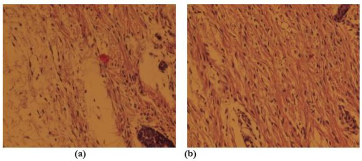
Fig. 2:

Microscopic view of open cutaneous wounds: (a) Control group, the number of Fibroblast are lesser than the experimental honey twice daily group in this photomicrograph. (b) Experimental honey twice-daily group, the number of Fibroblast is more than the control group in this photomicrograph. (Staining, H&E *40)
