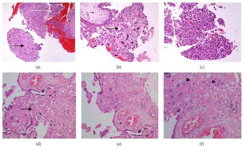
Figure 1.
Placental histological features from case 1 at 8 weeks of gestational age [(a), (b), and (c)] and case 2 at 32 weeks of gestational age [(d), (e), and (f)]. Chorionic villi (small arrow) surrounded by tumor trophoblast cells [cytotrophoblast and intermediate trophoblast with patchy small foci of syncytiotrophoblast (arrow)]. Mitotic figures (head arrow).

