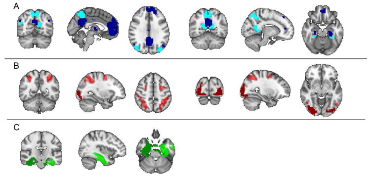
Figure 3.
Brain networks examined with resting-state fMRI analyses: Six networks based on Shirer et al. 2012 were selected due to their hypothesized recruitment by the memory task: (A) ventral (dark blue) and dorsal (light blue) default mode network, (B) higher visual (dark red) and visuospatial (light red) network, (C) left (dark green) and right (light green) medial temporal lobe.

