
Figure 4. Differential electrophysiological properties of Sst+ and Pvalb+ expressing EP neurons.
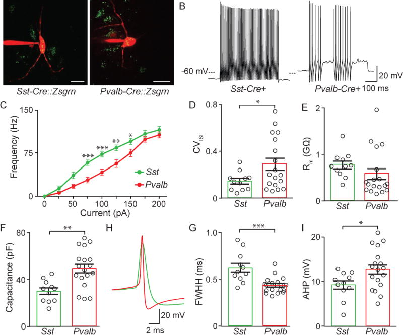
A. Images of Alexa Fluor-594 filled Zsgrn+ EP neurons in Sst-Cre and Pvalb-Cre mice crossed to Ai6 mouse.
B. Sample current-clamp recordings of action potential firing to +100 pA square wave current injection in Sst-Cre and Pvalb-Cre mice.
C. Action potential firing frequency vs current injection for neurons from Sst-Cre (green) (n=11 cells) and Pvalb-Cre (red) mice (n=18 cells).
D. The coefficient of variation of the interspike interval (CVISI) (averaged from ISIs across all current injections).
E, F. Membrane resistance (Rm) and capacitance across cell types.
G, H, I. Sample current-clamp recording of an action potential (G) and full width at half height (FWHH) (G) and after-hyperpolarization (AHP) (I) measurements from Sst-Cre (green) and Pvalb-Cre (red) neurons. All data are represented as mean ± SEM, *=p<0.05, **=p<0.01, ***=p<0.001. See also Figure S4 and S5.