
Figure 5. Sst+ EP neurons target LHb, and Pvalb+ neurons target LHb and motor thalamus.
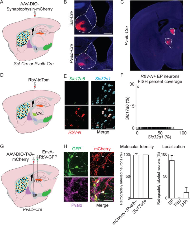
A. Illustration of a sagittal slice depicting AAV-DIO-Syn.-mCh. viral injection in EP in Sst-Cre or Pvalb-Cre mice.
B, C. Sample coronal image of axonal labeling (red) in the LHb (B) or VAL and AD thalamus (C) following viral injection into EP in Sst-Cre or Pvalb-Cre mice (DAPI in blue).
D. Illustration of a sagittal slice depicting RbV-tdTom viral injection in VAL.
E. Sample image of a coronal section of EP probed for RbV-N (red), Slc17a6 (green), and Slc32a1 (cyan).
F. Quantification of fluorescence coverage of Slc17a6 and Slc32a1 in RbV-N+ EP neurons (122 cells, n=3 animals).
G. Illustration of a sagittal slice depicting AAV-DIO-TVA-mCh. viral injection in EP, and EnvA-RbV-GFP injection in LHb in a Pvalb-Cre mouse.
H. (left) A sample image of a coronal section of EP showing RabV-GFP (green), mCh. (red), and Pvalb (magenta). (middle) Percentage of retrogradely labeled (GFP+) neurons that were also labeled for mCh. and Pvalb (169 cells, n=3 animals) or in separate FISH experiments Slc17a6 (25 cells, n=3 animals). (right) Quantification of soma location of GFP+ neurons following EnvA-RbV-GFP injection into LHb (186 cells, n=2 animals) (VAL=Ventral anterior lateral thalamus, AD=Anterior dorsal thalamus, LHA=Lateral hypothalamus, PF=Parafascicular nucleus of the thalamus, TRN=Thalamic reticular nucleus, MHb=medial habenula, V3=3rd ventricle). All data are represented as mean ± SEM. See also Figure S6.