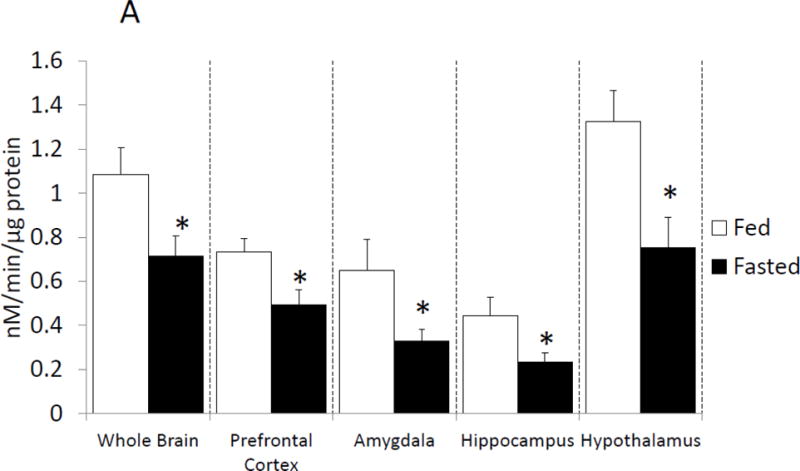
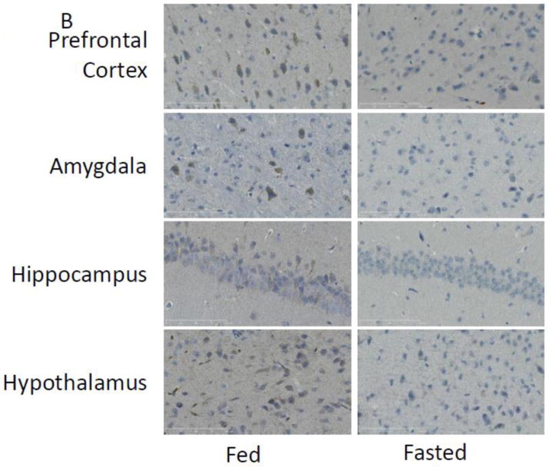
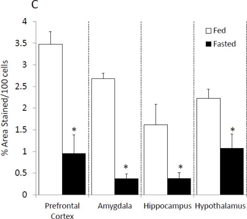
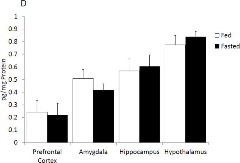
Figure. 1. Fasting reduces caspase-1 activity in the brain.


(A) Mice were given ad lib access to food (Fed) or fasted for 24 hours (Fasted). Caspase-1 activity was measured in whole brain and the brain regions indicated. Results are expressed as means ± SEM nM/min/μg total protein; n=7–10. (*p<0.05) (B) As in (A), mice were given ad lib access to food (Fed) or fasted for 24 hours (Fasted). Activated caspase-1 was examined in formalin-fixed paraffin embed sections by immunohistochemistry. Results are representative, 40X. (C) As in (A), mice were given ad lib access to food (Fed) or fasted for 24 hours (Fasted). As in (B), activated caspase-1 was examined in formalin-fixed paraffin embed sections by immunohistochemistry. Activated caspase-1 was quantified by image analysis. Results are expressed as means ± SEM % area stained/100 cells; n=4. (*p<0.05) (D) As in (A), mice were given ad lib access to food (Fed) or fasted for 24 hours (Fasted). IL-1β protein levels were measured by ELISA. Results are expressed as means ± SEM pg/mg Protein; n=7–10.
