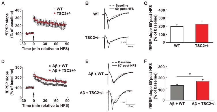
Fig. 6.
Aβ-induced hippocampal LTP impairments are alleviated by brain-specific deletion of TSC2. (A) HFS induced similar LTP in WT (open squares) and TSC2+/− (filled circles) hippocampal slices. Arrow indicates the time point at which HFS was delivered. (B) Representative fEPSP traces before and after HFS for LTP experiments shown in A. (C) Cumulative data showing mean fEPSP slopes 60 min after HFS based on the experiments shown in A. n = 7 for WT, n = 8 for TSC2+/−. unpaired independent t-test. p = 0.52. (D) Aβ-induced LTP impairments are mitigated in hippocampal slices derived from TSC2+/− mice (filled triangles) compared to WT slices (gray squares). Arrow indicates the time point at which HFS was delivered. (E) Representative fEPSP traces before and after HFS for the LTP experiments shown in D. (F) Cumulative data showing mean fEPSP slopes 60 min after HFS based on experiments shown in D. n = 9 for A+WT, n = 8 for Aβ+TSC2+/−. Unpaired independent t-test. *p < 0.05.

