Fig. 9.
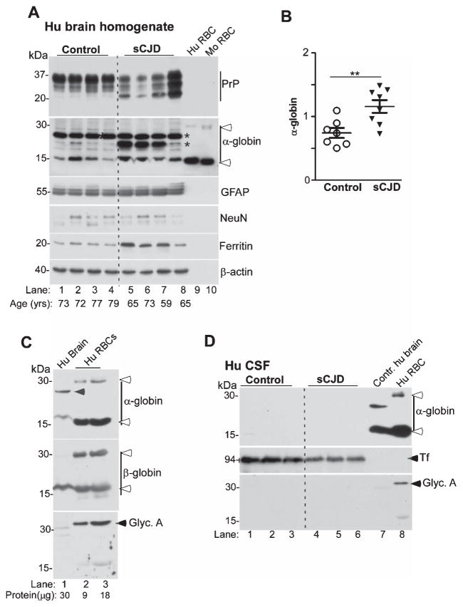
α-globin is increased in sCJD brains. A) Probing of lysates from the frontal cortex of sCJD and non-dementia controls shows significantly higher levels for α-globin in sCJD samples relative to controls (lanes 1–4 versus 5–8). Reaction for GFAP and ferritin is higher in sCJD, while NeuN is similar in control and sCJD samples (lanes 1–4 versus 5–8). Human and mouse RBCs fractionated in parallel confirm the presence of additional α-globin forms in human brain (lanes 1–8, * versus 9 & 10, open arrow-heads), and slower migration of monomeric α-globin from brain relative to RBC samples (lanes 1–8 versus 9 & 10). Reaction for β-actin provides a loading control. B) Quantification of α-globin by densitometry shows significantly higher levels in sCJD versus controls. C) Varying amounts of total protein from control human brain and human RBCs fractionated in parallel shows a strong reaction for α-globin and β-globin in brain and RBC samples (lanes 1–3, top and middle panels). A faster migrating form of brain α-globin that does not co-migrate with dimeric β-globin from RBCs is detected as in Figs. 7 and 8 above (lane 1, top panel, arrow-head). Reaction for glycophorin-A is limited to RBC samples (lanes 2 & 3, bottom panel), ruling out contamination of brain samples with RBCs. D) Western blotting of equal volume of CSF from sCJD and control samples shows no reactivity for α-globin, though Tf is detected readily, and is reduced in sCJD samples (lanes 1–3 versus 4–6) [61]. Human brain and RBC samples react readily for α-globin as expected (lanes 7 & 8). Reaction for glycophorin-A is limited to the RBC sample (lane 8).

