. 2017 Mar 31;8(5):4100–4107. doi: 10.1039/c7sc00646b

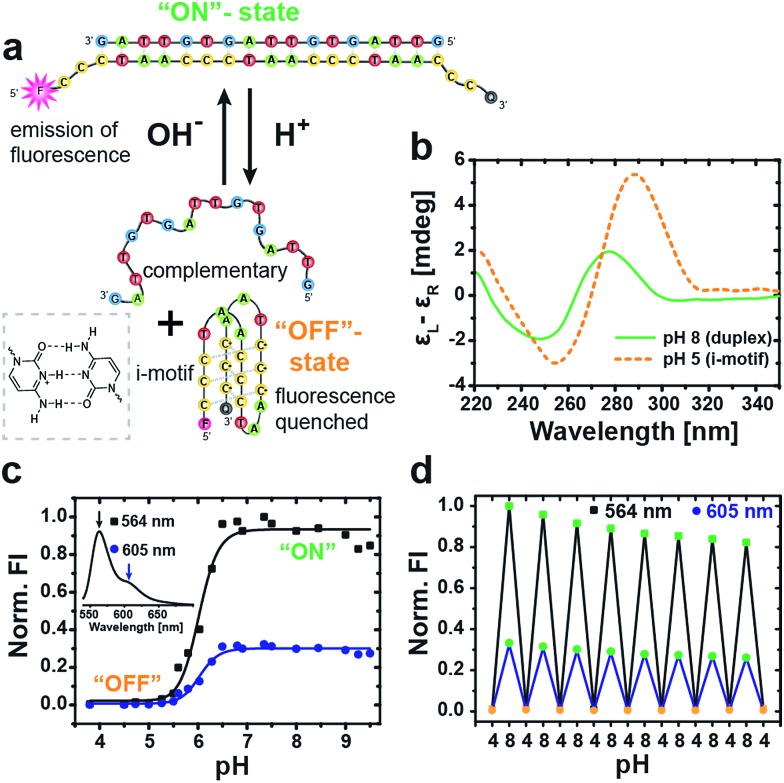
Fig. 1. Characterization of the i-motif switch under different static pH conditions. (a) Schematic illustration of the two conformational states of the i-motif switch with the closed hybridized “on”-state (duplex) at alkaline pH and the open “off”-state (i-motif + random coil) at acidic pH. The i-motif tetraplex is stabilized by hemiprotonated cytosine+–H–cytosine base pairs (left box). The i-motif switch is modified with a Cy3 fluorophore (F) and a Black Hole Quencher 2 (Q) to produce fluorescence upon photoexcitation (λ exc = 520 nm) as output signal for the active “on”-state at high pH. (b) CD spectra of the unlabeled switch show the characteristic bands of duplex B-DNA at pH 8 and of the folded i-motif at pH 5. (c) Fluorescence intensity (FI, normalized to maximum) of the i-motif switch in dependence of pH evaluated at the two maxima of the Cy3 fluorophore at 564 nm (black squares) and 605 nm (blue dots). Inset shows the full emission spectrum of Cy3 for the activated i-motif switch (“on”-state). (d) Multiple cycling between acidic and alkaline pH switches the fluorescence reversibly “on” and “off”. Slight dilution effects due to addition of acid and base are visible.

Fig. 1