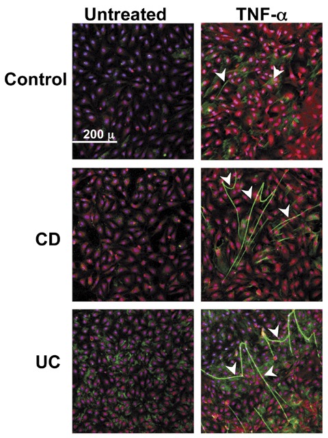
Figure 3.

HA deposition and cable formation is increased in vitro on TNF‐α‐stimulated intestinal EC. Immunohistochemistry of three isolates of HIMEC that were treated without or with TNF‐α (10 ng/mL) for 18 hours and specifically stained for HA (green), von Willebrand Factor (red), and nuclei (blue). Arrowheads call attention to the cable‐like HA structures on the HIMEC surfaces that can span multiple cell lengths. The figure shown is representative of three or more different patient HIMEC isolates per patient category.
