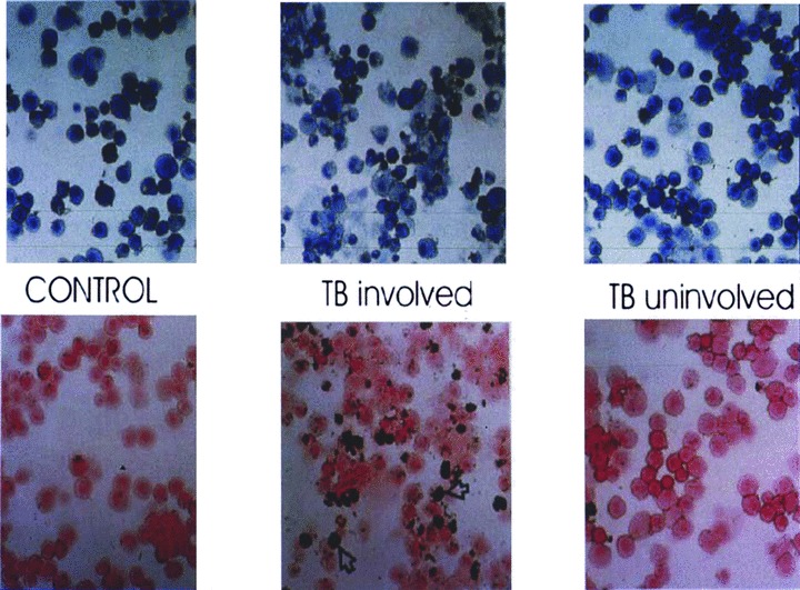
Figure 2.

Giemsa (upper panel) and peroxidase (lower panel) stains of bronchoalveolar lavage (BAL) from TB patient and control. BAL from the involved segment shows lymphocytic alveolitis. Approximately 40% of the bronchoalveolar cells are peroxidase positive moncyte‐derived immature macrophages. Reprinted from Reference 22 with permission.
