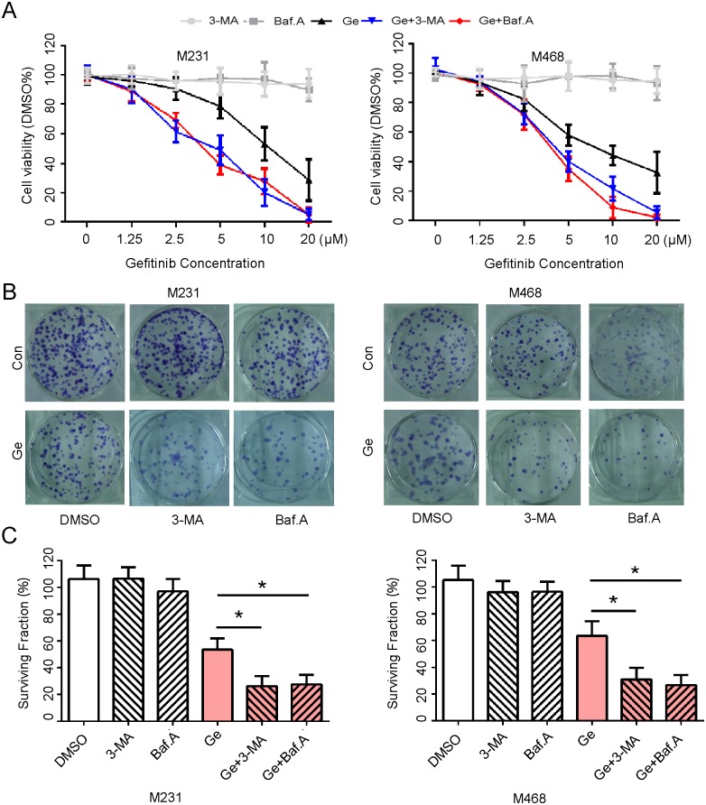
Fig 2. Autophagy inhibitor facilitates cytotoxicity of Ge in TNBC cells in vitro.
(A) MDA-MB-231 and MDA-MB-468 cells were treated with 0, 1.25, 2.50, 5.00, 10.00, 20.00 μM Ge alone or combined with 3-MA (10 mM) or Baf.A (1 nM) respectively for 48 hours, DMSO acted as the control, and then subjected to CCK8 assay. Absorbance value was calculated and standardized to DMSO group. Three independent experiments were performed. (B) The above cells were treated with DMSO (0.2%), 3-MA (10 mM), Baf.A (1 nM), Ge (5 μM), Ge (5 μM) +3-MA (10 mM) and Ge (5 μM) +Baf.A (1 nM), DMSO acted as the control, and subjected to cell colony formation assay. (C) Cell surviving fraction were calculated and presented as mean ± SD; *p < 0.05. Three independent experiments were performed.

