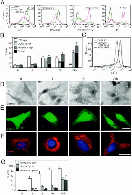
FIG. 1.
HCQ-mediated induction of acidic vacuoles. (A and B) Vacuolar acidic compartment- and apoptosis-associated parameters in HCQ-treated cells. HeLa cells were exposed to HCQ (30 μg/ml) for the indicated periods, and cells were stained with LTR, DiOC6(3), annexin V-fluorescein isothiocyanate, or PI, followed by FACS analysis. Data shown are representative FACS profiles (A) or means of results from five independent experiments (x ± standard errors of the mean [SEM]) (B). Bars in panel A indicate the window representing each population. CRT, control. (C) Effect of mitochondrion-stabilizing proteins on LTR staining. HeLa cells stably transfected with Bcl-2 or vMIA were incubated for 5 h with HCQ, followed by LTR staining and FACS analysis. Vector-only control cells (unpublished results) behaved as cells for which results are shown in the leftmost graph of panel A. (D and E) Light microscopic evidence for HCQ-induced vacuolization. Cells treated with HCQ for the indicated periods were stained with Giemsa (D) or Cell Tracker Green CMFDA (E). The arrow indicates the apoptotic nucleus. (F) Staining of lysosomes with a LAMP2 antibody. HCQ-treated HeLa cells were immunofluorescence stained and counterstained with Hoechst 33324. (G) Chronological hierarchy of vacuolization and MMP. Cells were stained with CMFDA, an anti-cytochrome c (Cyt c) antibody (revealed as red fluorescence), and Hoechst 33342 (blue fluorescence), and the frequencies of cells with enhanced vacuolization, mitochondrion-released cytochrome c, and apoptotic nuclei were determined (x ± SEM; n = 4).

