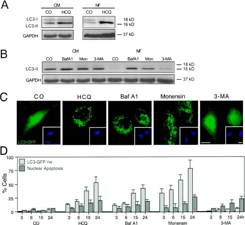
FIG. 4.
Effect of HCQ and other autophagy inhibitors on the subcellular localization and biochemical status of the autophagic vacuole marker LC3. (A and B) Immunoblot analyses of accumulating LC3-II protein in control (CM) and starved (NF, 6 h) cells treated with HCQ (A) or a range of established autophagy inhibitors (B). (C and D) Redistribution of LC3-GFP. Twenty-four hours after transient transfection with an LC3-GFP chimera, cells were treated for the indicated times (24 h in panels C in the presence of serum) with HCQ, Baf A1, monensin, or 3-MA; fixed; and counterstained with Hoechst 33342. Representative cells are shown in panels C, and the frequency (x ± SEM; n = 4) of cells with a clear vacuolar distribution of LC3-GFP (LC3-GFPVac) or apoptotic nuclei was scored. CO, control; Mon, monensin.

