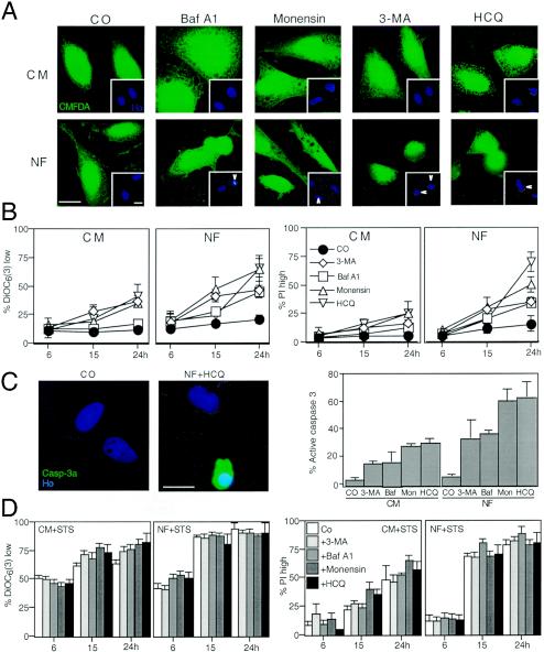
FIG. 5.
Autophagy inhibition sensitizes cells to nutrient depletion-induced cell death. (A) Vacuolization induced by starvation plus autophagy inhibition. HeLa cells were cultured in CM or NF for 24 h in the presence or absence (control [CO]) of HCQ, Baf A1, monensin, or 3-MA and finally stained with CMFDA and Hoechst 33342 (inset). Note that HCQ, Baf A1, and monensin enhance the formation of AV (visible as holes in the green fluorescent staining) and induce nuclear apoptosis. Arrows mark apoptotic nuclei. (B) Quantitative assessment of synergic cell death induction. Cells cultured in CM or NF, in the presence of the indicated inhibitors or none of them (control), were stained to determine the loss of ΔΨm [with DiOC6(3)] and viability (with PI). Data shown are means of results of five independent experiments ± SEM. (C) Caspase-3 activation (Casp-3a) triggered by nutrient depletion plus autophagy inhibition. Cells were stained with an antibody recognizing the 17-kDa subunit of activecaspase-3, and the frequency of positive cells was scored after culture in the presence or absence of nutrient and autophagy inhibitors (as described for panel B, at 24 h). Ho, Hoechst 33342; Baf, Baf A1; Mon, monensin. (D) Autophagy inhibition does not sensitize cells to STS-induced cell death. Cells were cultured with 100 nM STS in the presence of the indicated inhibitors, and cell death-related parameters were measured as described for panels B. Results are mean values ± SEM of three to five independent determinations.

