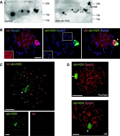
FIG. 1.
ubi-H2A marks the XY bodies and telomeres of pachytene spermatocytes. A: Western blots of basic nuclear proteins separated on two-dimensional AUT-SDS-polyacrylamide gels (first dimension, AUT; second dimension, SDS) were stained with anti-ubi-H2A (right panel) or with second antibody only (control, left panel). A specific signal is detected at the expected position for ubi-H2A (arrowhead). The approximate positions of histones H2A, H2B, tH3, and H4 are indicated for reference. Positions and molecular weights of marker proteins are indicated. The expected approximate position of ubi-H2B is indicated by the white ellipse, and the blots are overexposed to show that no ubi-H2B is detected. B: Spermatocyte spread nuclei from wild-type mice stained with a mouse monoclonal IgG antibody against Sycp3 (blue), a rabbit polyclonal antiubiquitin antibody (red), and a mouse monoclonal IgM antibody against ubi-H2A (green). Both ubi and ubi-H2A mark XY body chromatin (arrowhead in right panel, showing the merged signals). Anti-ubi also detects other ubiquitinated proteins associated with chromatin in the rest of the nucleus. Upon overexposure, ubi and ubi-H2A are detected at the ends of some synaptonemal complex axes (insets). C: FISH analysis of telomeres (tel) (red) followed by immunocytochemistry with anti-ubi-H2A (green) of an early diplotene cell. Diplotene is identified by the presence of a low level of ubi-H2A in the XY body (arrowhead) and the high number of telomeric foci (more than 40, with double foci still attached counted as one) and splitting of telomeres. The upper panel shows that around 30 telomere foci colocalize, at least partially, with ubi-H2A. A total of approximately 100 ubi-H2A foci are observed outside the XY body area, indicating that chromatin regions other than the XY body and (near) telomeric regions also accumulate ubi-H2A. The bottom panels show lower-magnification images of the single fluorescent signals. D: Spermatocyte spread nuclei from rat and human stained with anti-Sycp3 (red) and anti-ubi-H2A (green). ubi-H2A marks XY body chromatin (arrowhead) in both species. Bars, 20 μm.

