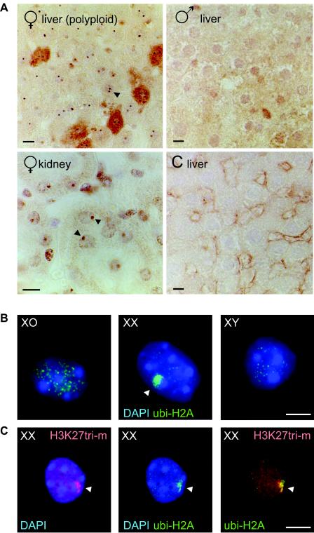
FIG. 2.
ubi-H2A marks the inactive X chromosome (Barr body) in female somatic cells. A: Immunohistochemical analysis of ubi-H2A localization in rat female liver and kidney sections and in male liver sections. Control (C liver): incubation without first antibody. In female kidney nuclei, high-level accumulation of ubi-H2A is seen as a single nuclear spot (arrowhead). In addition, two spots are often observed in polyploid female liver nuclei (arrowhead). These spots are not observed in male tissues. Bars, 10 μm. B: Immunocytochemical detection of ubi-H2A in XO, XX, and XYtdym1 pregranulosa cells. A variable number of small foci, of unknown nature, is present in all nuclei, but only the XX pregranulosa cells accumulate ubi-H2A in a single large spot in the periphery of the nucleus (arrowhead). Bar, 20 μm. C: Immunocytochemical detection of H3 lysine 27 trimethylation (H3K27tri-m) (red) and ubi-H2A (green) in XX pregranulosa cells. H3 lysine 27 methylation and ubi-H2A largely colocalize in a single large spot in the periphery of the nucleus (arrowhead). Bar, 20 μm.

