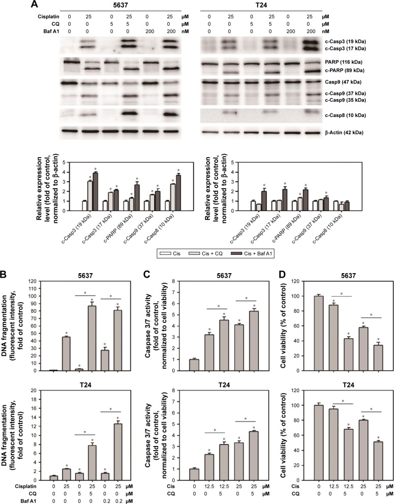
Figure 4.
CQ and Baf A1 enhanced cisplatin-induced apoptosis.
Notes: (A) The expression levels of proapoptotic marker proteins, (B) DNA fragmentation, (C) caspase 3/7 activities, and (D) cell viability in cells treated with DMSO or 25 µM cisplatin with or without 2 hours pretreatment of either 5 µM CQ or 200 nM Baf A1 for 24 hours. The expression level of c-Casp3, c-PARP, c-Casp9, and c-Casp8 was detected by Western blot. The expression of β-actin served as internal control. Representative blots from three independent experiments are shown. The relative band intensities were quantitated by densitometric scanning and the relative expression levels are presented as the fold of control cells (lower panels). The values in (B–D) are shown as the mean ± SD of three independent experiments; *P<0.05.
Abbreviations: SD, standard deviation; DMSO, dimethyl sulfoxide; Baf A1, bafilomycin A1; CQ, chloroquine; c-PARP, cleaved poly(adenosine diphosphate ribose) polymerase; c-Casp, cleaved caspase; Cis, cisplatin.

