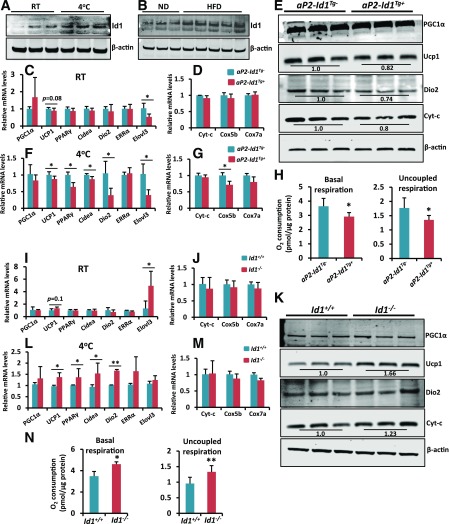
Figure 4.
Thermogenic protein levels and VO2 rate are reduced in the BAT of aP2-Id1Tg+ mice. A: Expression levels of Id1 and β-actin in the BAT of 2-month-old wild-type mice at RT and after exposing mice to 4°C for 4 h. B: Expression levels of Id1 and β-actin in the BAT of 2-month-old ND- or HFD-fed (1 week) wild-type mice. C and D: Relative mRNA transcript levels of thermogenic and mitochondrial genes in the BAT of aP2-Id1Tg− and aP2-Id1Tg+ male mice at RT. E: Expression levels of indicated proteins in the BAT of 2-month-old aP2-Id1Tg− and aP2-Id1Tg+ male mice at RT. F and G: Relative mRNA transcript levels of thermogenic and mitochondrial genes in the BAT of aP2-Id1Tg− and aP2-Id1Tg+ male mice after exposure to 4°C for 4 h. H: Basal respiration and uncoupled respiration (after blocking ATP synthase with oligomycin) were determined in the BAT explants of aP2-Id1Tg− and aP2-Id1Tg+ male mice with an Oxygraph Plus System (n = 4). I and J: Relative mRNA transcript levels of thermogenic and mitochondrial genes in the BAT of Id1+/+ and Id1−/− male mice at RT. K: Expression levels of indicated proteins in the BAT of 2-month-old Id1+/+ and Id1−/− mice at RT. After acquiring images, band intensities were measured and normalized to β-actin by the Li-Cor Odyssey system. L and M: Relative mRNA transcript levels of thermogenic and mitochondrial genes in the BAT of Id1+/+ and Id1−/− male mice after exposure to 4°C for 4 h (n = 6–8). N: Basal respiration and uncoupled respiration were determined in the BAT explants of Id1+/+ and Id1−/− mice (n = 4). Data are mean ± SD. *P < 0.05; **P < 0.005.

