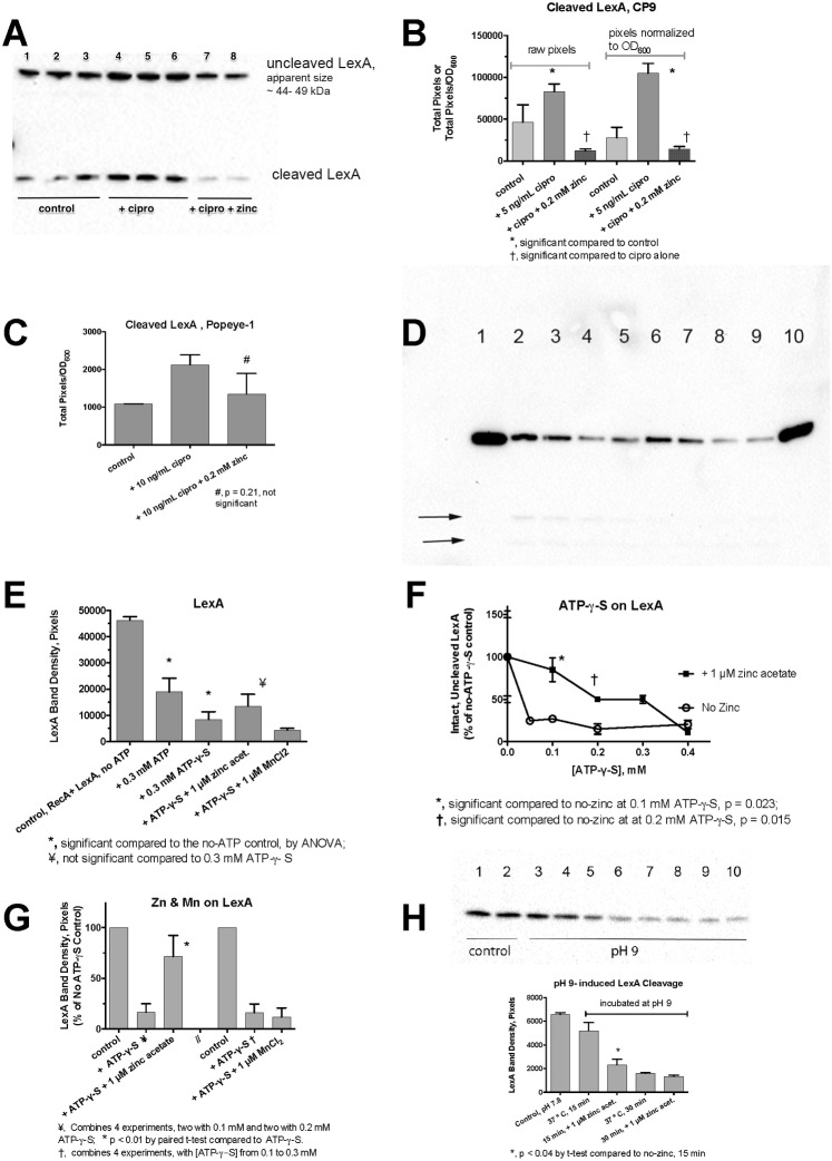
Fig 5. Regulation LexA by SOS activators and by zinc in E. coli.
Panel A, immunoblot for LexA in whole-cell extracts of cultures of E. coli CP9 after a 3 h exposure to ciprofloxacin with and without zinc. Uncleaved LexA appeared to migrate in the form of a LexA dimer in these blots; while the cleaved LexA product ran at ~ 15 kDa. Panel B, densitometry scan of blot in Panel A, raw (left) and corrected for the effects of treatment on growth (right panel). Panel C, densitometry scan of a LexA blot (not shown) after a 1 h exposure to ciprofloxacin in Popeye-1. Panels D- H, RecA-mediated LexA cleavage assays in vitro, showing immunoblots against LexA. Purified LexA and RecA were incubated in vitro in the presence of absence of necessary cofactors, such as ssDNA and ATP or ATP- γ -S as described in the Methods section Panel D, RecA-mediated cleavage of LexA. An unlabeled lane to the left of lane 1 contained RecA alone, showing that the antibody does not cross-react between the two proteins. All the labeled lanes in Panel D received RecA, LexA, and a 38-mer oligonucleotide. Lane 1, no ATP; Lanes 2 and 3 also received 0.3 mM ATP; Lanes 4 and 5 also received 0.3 mM ATP-γ-S. Faint LexA cleavage products were visible in lanes 2–5 in the original blots, arrows; Lanes 6 and 7, plus ATP-γ-S and 1 μM zinc acetate; Lanes 8 and 9, plus ATP-γ-S and 1 μM MnCl2; Lane 10 received 0.3 mM GTP, which does not support RecA activation, as an additional control. Panel E, densitometry scan of the chemiluminescence signal from the blot shown in Panel D. Panel F, dose-response relationship of ATP-γ-S concentration vs. LexA cleavage in the absence and presence of 1 μM zinc acetate, showing protection by zinc against LexA cleavage at 0.1 to 0.3 mM ATP-γ-S. Panel G, combined results of 4 separate experiments testing for the effect of zinc acetate, and four experiments with MnCl2 on LexA cleavage, with results normalized to the no- ATP-γ-S control so that separate experiments could be compared. Panel H, lack of protection by zinc on LexA auto-cleavage induced by incubation at pH 9. Control lanes 1 and 2 show LexA kept at pH 7.8; Lanes 3–6 show LexA protein incubated for 15 min at pH 9, 37°. Lanes 7–10 show samples incubated at pH 9 for 30 min, 37°. Lanes 5–6 and 9–10 also received 1 μM zinc acetate.

