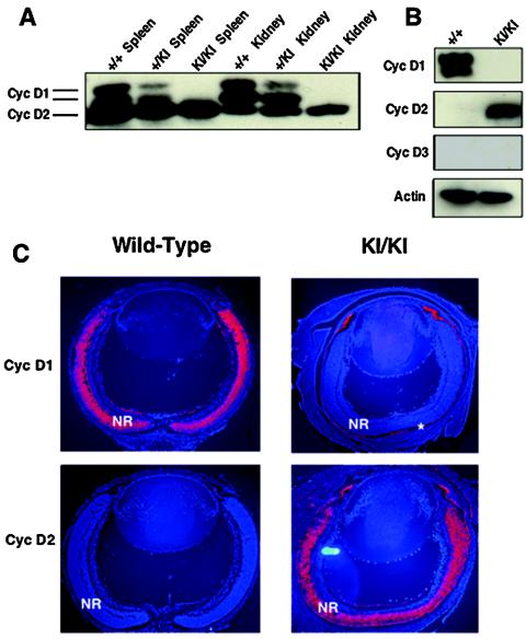
FIG. 2.
Molecular analyses of cyclin D2→D1 tissues. (A) Western blot analysis of indicated organs from wild-type (+/+), heterozygous (+/KI), or homozygous cyclin D2→D1 mice (KI/KI) probed with an antibody against cyclins D1 and D2. (B) Western blot analyses of retinas dissected from 1-day-old pups, probed with antibodies against cyclin D1, D2 or D3 or actin (loading control). (C) In situ hybridization analyses of retinas dissected from 1-day-old wild-type or cyclin D2→D1 (KI/KI) mice. Sections were hybridized with cyclin D1 or D2 cDNA probes. Red coloring represents the positive hybridization signal. Blue coloring represents counterstaining of cell nuclei with Hoechst stain. NR, neuroretina. The positive staining of the pigment cell layer (asterisk) represents an artifact, as this layer frequently stains with all probes.

