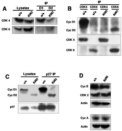
FIG. 3.
Molecular analyses of cyclin D2→D1 retinas. (A) Protein lysates isolated from postnatal day 1 retinas were immunoprecipitated with an anti-cyclin D1 (+/+, wild-type samples) or anti-cyclin D2 antibodies (KI/KI, cyclin D2→D1 samples), followed by immunoblotting with anti-CDK4 or anti-CDK6 antibodies. The left panel (Lysates) shows immunoblots of straight lysates probed with antibodies against CDK4 or CDK6. (B) Retinal lysates were immunoprecipitated with antibodies against CDK4 or CDK6, followed by immunoblotting with an antibody recognizing cyclins D1 and D2 or with anti-CDK4 or anti-CDK6 antibodies. (C) Retinal lysates were immunoprecipitated with an antibody against p27Kip1. Immunoblots were probed with an antibody recognizing cyclins D1 and D2. The left panel (Lysates) shows immunoblots of straight lysates probed with antibodies against cyclin D1 and D2 or against p27Kip1. (D) Western blot analyses of retinal lysates probed with the indicated antibodies. Antiactin antibodies were used to ensure equal loading.

