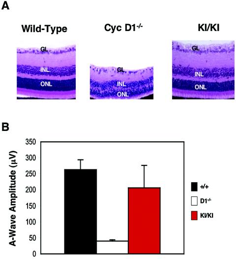
FIG. 4.
Rescue of the retinal abnormalities. (A) Hematoxylin and eosin-stained sections of retinas collected from 3-week-old wild-type, cyclin D1−/−, or cyclin D2→D1 (KI/KI) animals. GL, ganglion cell layer; INL, inner nuclear layer; ONL, outer nuclear layer. Magnification, ×40. (B) Mean amplitudes of a-waves generated in the retinas in response to a pulse of light (ERG testing). Error bars indicate standard deviations.

