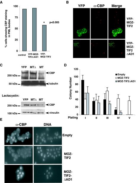
FIG. 6.
Effect of MOZ-TIF2 on cellular CBP protein levels and on the proliferation of hematopoietic stem cells in vitro. (A) Quantitative analysis of CBP-containing speckles (PML bodies) in control cells (n = 300) and YFP-MOZ-TIF2- and YFP-MOZ-TIF2ΔAD1-expressing cells (n = 50). (B) Representative images of COS-1 cells, showing the effect of YFP-MOZ-TIF2 and YFP-MOZ-TIF2ΔAD1 expression on detection of endogenous CBP speckles. (C) Western blot analysis of whole-cell extracts from YFP+ sorted HEK293 cells expressing YFP, YFP-MOZ-TIF2,and YFP-MOZ-TIF2ΔAD1 cultured in the absence (upper panels) or presence (lower panels) of lactacystin. The band corresponding to full-length CBP is indicated. The loading controls α-tubulin and vinculin are also indicated. (D) Serial replating assays to assess the proliferative potential of GFP+ Lin− cells transduced with the indicated vectors in methylcellulose medium containing SCF, IL-3, and Il-6. Error bars indicate standard errors of the means. (E) Anti-CBP staining of sorted GFP+ Lin− cells transduced with the indicated retroviral vectors. Images were taken at identical exposure times. DNA was counterstained with 0.1 μg of DAPI per ml.

