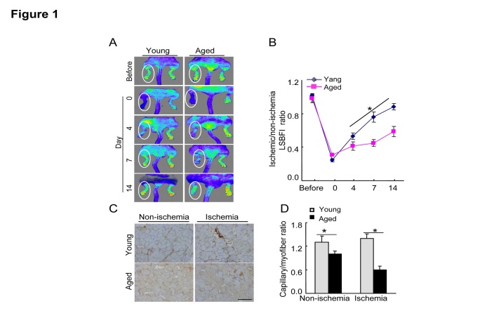
Figure 1. Aging impaired blood flow recovery and capillary formation in ischemic tissues.
A) Serial LSBFI showed that ischemic hindlimbs of the aged mice (>18 mos-old) exhibited a low perfusion signal (dark blue), whereas those of the young (2-mos-old) mice exhibited a high signal (red). B) The ratio of ischemia to normal LSBGI was lower in the aged mice compared to the young mice. C) On postoperative day 14, immunostaining was conducted to evaluate the capillaries in non-ischemic and ischemic thigh adductor muscles. D) Quantitative analyses revealed that aging reduced the capillary density in both non-ischemic and ischemic muscle compared to young mice. Data are mean ± SEM (n=5-6). *P<0.05 vs. corresponding controls (day 4, 7, and 14) by two-way repeated-measures ANOVA and Bonferroni post hoc tests or one-way ANOVA and Tukey’s post hoc tests. Scale bar, 50 μm.

