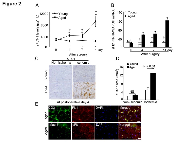
Figure 2. Aging increased the plasma and the ischemic muscle sFlt1 levels.
A) The ELISA showed that the aged mice had higher levels of circulating sFlt1 protein throughout the follow-up period (n=6-8). B) Quantitative real-time PCR showed that the aged mice had higher levels of sFlt1 gene throughout the follow-up period (n=6-8). Data are mean ± SEM. *P<0.05 vs. corresponding day 0; †P<0.05 vs. corresponding young mice during ischemia; two-way ANOVA and Bonferroni post hoc tests. C and D) Representative images and quantitative data shows that sFlt-1+ (including splice isoform sFlt-1 [77 kDa] and sFlt1 isoform 14 [82 kDa]) staining signal was markedly increased in the ischemic myofiber space of aged mice at postoperative day 4. E) Representative triple imunofluorescent images show that Flt-1 is expressed in endothelial cells as well as macrophages. Data are mean ± SEM (n=6). *P<0.05, †P<0.05 by one-way ANOVA and Tukey’s post hoc tests. NS indicates no significant. Scale bar, 50 μm.

