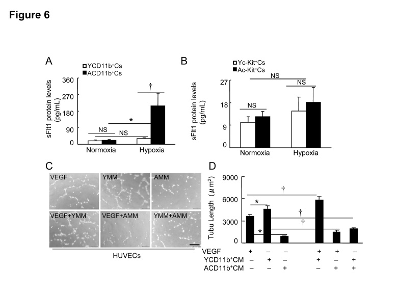
Figure 6. The effects of hypoxic stress on sFlt1 production in BM-derived c-Kit+ cells and CD11b+ cells of young and aged mice.
A and B) Subconfluent young mouse BM-derived c-Kit+ cells (Yc-Kit+Cs) or CD11b+ cells (YCD11b+Cs) and aged mouse BM-derived c-Kit+ cells (Ac-Kit+Cs) or CD11b+ cells (ACD11b+Cs) were cultured (six-well-plates) in serum-free EBM-2 (for the former) or RPMI medium 1640 (for the later) under hypoxic condition (plates in hypoxic cambers) for 36 hr (for c-Kit+ cells) or 48 hr (CD11b+ cells), respectively, and the conditioned media were then subjected to the ELISA with sFlt1 kits. C and D) Following collection of the culture medium (Yc-Kit+CM, Ac-Kit+CM; YCD11b+CM, ACD11b+CM) as the same as above, the special fraction containing an approx. >70 kDa protein was isolated with 150 K and then 50 K AmiconUltra contricons, and we adjusted the protein concentration to 1.5 mg/ml for the cellular experiments. HUVECs (2 × 10⁴) were cultured (24-well-plates) in EBM-2 containing VEGF-A, Yc-Kit+CM, Ac-Kit+CM, VEGF/Yc-Kit+CM, VEGF/Ac-Kit+CM, or Yc-Kit+CM/Ac-Kit+CM (20 ng/mL for VEGF-A; 90 μg/mL for the cultured media) respectively for 24 hr, and then subjected to length calculation. Representative images (C) and combined data (D) show the tubulogenesis in response to each stimulator. Data are mean ± SEM (n=6~8). *P<0.05 by one-way ANOVA and Tukey’s post hoc tests. Scale bar, 50 μm.

