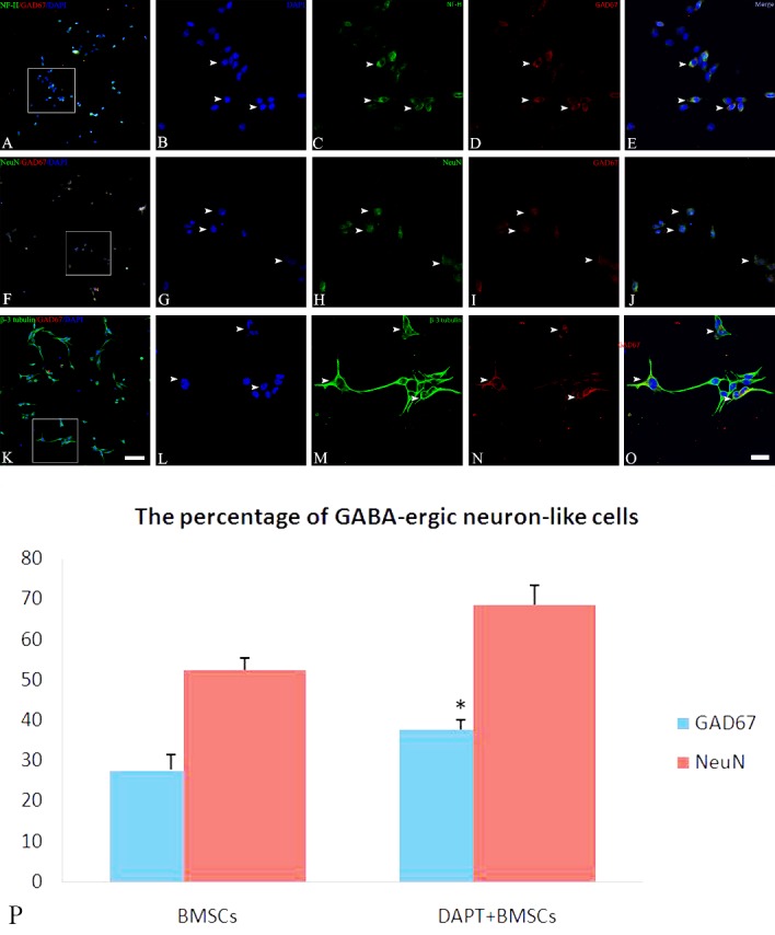
Figure 4. Immunofluorescence for GABA-ergic neuron-like differentiation from BMSCs.

After BMSCs were induced by the cocktail conditioned medium, immunofluorescence showed that differentiated cells expressed neuronal cytoskeleton marker NF-H (C, white arrow), mature neuronal marker NeuN (H, white arrow), specific neuronal marker (β-3 tubulin (M, white arrow), and GABA-ergic cell marker GAD67 (D, I and N, white arrow). In addition, a few GAD67 positive cells are co-localized with NF-H (A / E, white arrow), NeuN (F/J, white arrow), and β-3 tubulin (K/O, white arrow). B, G and L illustrate DAPI stained nuclei. Bar (A, F and K) = 50 μm, bar (E, J and O) = 20 μm. Statistical analysis shows the percentage of GABA-ergic neuron-like differentiation in DAPT treated BMSCs (P). *P = 0.002, < 0.01, vs. BMSCs.
