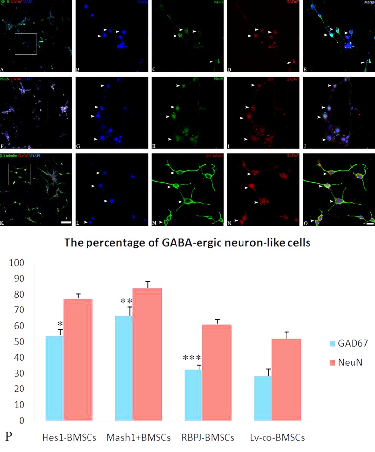
Figure 5. Immunofluorescence showing GABAergic neuron-like differentiation from Mash1 overexpressed BMSCs.

After Mash1+ BMSCs were induced by cocktail conditioned medium, immunofluorescence showed that differentiated cells expressed neuronal cytoskeleton marker NF-H (C, white arrow), mature neuronal marker NeuN (H, white arrow), and specific neuronal marker β-3 tubulin (M, white arrow), in addition to GABA-ergic cell marker GAD67 (D, I and N, white arrow). Note that, many GAD67 positive cells co-localize with NF-H (A / E, white arrow), NeuN (F / J, white arrow), and β-3 tubulin (K/O, white arrow). B, G and L illustrate DAPI stained nuclei. Bar (A, F and K) = 50 μm, bar (E, J and O) = 20 μm. Statistical analysis shows the percentage of GABA-ergic neuron-like differentiation in genetically engineered BMSCs (P). Statistical differences referred to Lv-con-BMSCs, *P = 1.9036E-05, < 0.01; **P = 3.61045E-05, < 0.01; ***P = 0.122, > 0.05.
