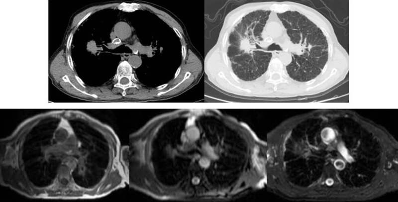
Figure 3.

Phase I of CWP complicated by a large shadow formation, with CT scanning (figure above) suggesting the possibility of pneumoconiosis to form lung cancer. MRI showing tumors in equal-low signal shadows, consistent with the representations of large shadows of pneumoconiosis. Diagnosis confirmed after 2 years of follow-up. CT = computed tomography, CWP = coal worker pneumoconiosis, MRI = magnetic resonance imaging.
