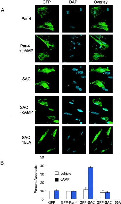
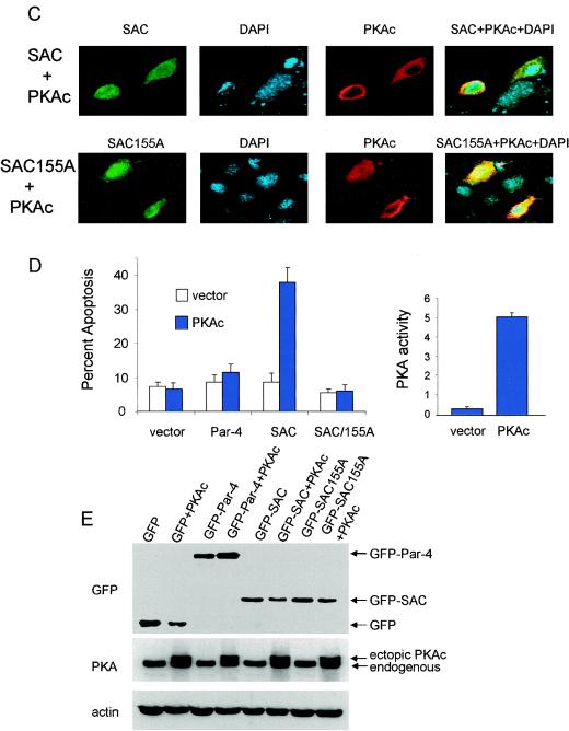
FIG. 4.
Elevation of PKA activity in normal cells activates the apoptotic potential of the SAC domain of Par-4 in a T155-dependent manner. MEF cells were transiently transfected with vector, GFP-Par-4, GFP-SAC, or GFP-SAC/155A expression constructs, treated with either vehicle or 8-Cl-cAMP (10 μM) for 48 h, and examined for intracellular localization of Par-4 or mutants (A) or apoptosis (B). MEF cells were cotransfected with expression constructs for GFP-Par-4, GFP-SAC, or vector and PKAc for 48 h; the transfected cells were visualized under a confocal microscope by GFP fluorescence for Par-4 or mutants or by immunostaining with PKAc antibody, followed by Texas red-conjugated secondary antibody, and for apoptosis by DAPI staining (C). Apoptotic cells were scored and the data were presented as percent apoptosis (D, left). PKA activity was determined with the cAMP-dependent PKA Signatect assay kit (D, right). Protein expression was examined by Western blot analysis with antibodies for GFP, PKAc, or actin (E).


